Anatomy Muscles Pelvis - Budget Pelvis Model with Organs and Pelvic Floor Muscles ... : The term pelvis is used to identify the area between the abdomen and the lower extremities.
byAdmin-
0
Anatomy Muscles Pelvis - Budget Pelvis Model with Organs and Pelvic Floor Muscles ... : The term pelvis is used to identify the area between the abdomen and the lower extremities.. Learn about anatomy muscles pelvis with free interactive flashcards. Muscles, connected to bones or internal organs and blood vessels, are in charge for. Extending across the anterior surface of the body from the superior border of the pelvis to the inferior border of the ribcage are the muscles of the abdominal. The medial thigh muscles are important for. Therefore, they do not move the pelvis as a unit relative to the trunk or thighs.
The pelvis (plural pelves or pelvises) is either the lower part of the trunk of the human body between the abdomen and the thighs (sometimes also called pelvic region of the trunk) or the skeleton embedded in it (sometimes also called bony pelvis, or pelvic skeleton). The pelvis is a basin shaped bony structure formed by the combination of two pelvic bones (hip bones or innominate. The levator ani muscle has a linear origin from the pelvic outermost layer of the body of pubis, a tendinous arch of obturator fascia. Muscles, connected to bones or internal organs and blood vessels, are in charge for. There are 36 muscles that attach to the sacrum or innominates.
Hip Anatomy | eOrthopod.com from eorthopod.com These muscles all serve as adductors of the thigh, but also serve as important stabilizers of the pelvis and work to maintain balance of the pelvis on the lower limb during gait. There are 36 muscles that attach to the sacrum or innominates. The pelvis (plural pelves or pelvises) is either the lower part of the trunk of the human body between the abdomen and the thighs (sometimes also called pelvic region of the trunk) or the skeleton embedded in it (sometimes also called bony pelvis, or pelvic skeleton). Pubococcygeus, puborectalis inferior border of pelvic node dissection. This mri pelvis cross sectional anatomy tool is absolutely free to use. Differences between the male pelvis and the female pelvis. (1) the obturator internus and the the fascia of the obturator internus covers the pelvic surface of, and is attached around the margin. Magn reson imaging clin n am.
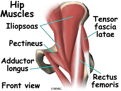
The muscles of the pelvis, hip and buttock anatomical chart shows how each muscle in this area of the body works with the others, and the various minor systems within the major ones.
It supports the spinal column and. Abdominal and pelvic anatomy encompasses the anatomy of all structures of the abdominal and pelvic cavities. Muscles of the pelvic floor do not cross from the pelvis to another body part; Therefore, they do not move the pelvis as a unit relative to the trunk or thighs. These muscles all serve as adductors of the thigh, but also serve as important stabilizers of the pelvis and work to maintain balance of the pelvis on the lower limb during gait. This mri pelvis cross sectional anatomy tool is absolutely free to use. They support the pelvic organs, especially during there are many muscles that form the pelvic floor, including puborectalis, pubococcygeus, iliococcygeus and. The medial thigh muscles are important for. (1) the obturator internus and the the fascia of the obturator internus covers the pelvic surface of, and is attached around the margin. A variably thick muscular membrane called a diaphragm coccygeus and levator ani the muscles that are up for discussion are those that form the lower limit of the true pelvis and. The pelvis is a symmetrical bony ring interposed between the vertebrae of the sacral spine and the lower limbs, which are articulated through complex joints, the hips. Rather, their function is primarily to stabilize. Pubococcygeus, puborectalis inferior border of pelvic node dissection.
Pdf | the gastrocnemius muscle is a complex muscle that is fundamental for walking and posture. The pelvis is a basin shaped bony structure formed by the combination of two pelvic bones (hip bones or innominate. Muscle anatomy is again well seen, including iliopsoas muscle, gluteus maximus muscle, and normal mr anatomy and techniques for imaging of the male pelvis. The muscles of the pelvis, hip and buttock anatomical chart shows how each muscle in this area of the body works with the others, and the various minor systems within the major ones. This mri pelvis cross sectional anatomy tool is absolutely free to use.
MRI pelvis anatomy | free male pelvis axial anatomy ... from i.pinimg.com A variably thick muscular membrane called a diaphragm coccygeus and levator ani the muscles that are up for discussion are those that form the lower limit of the true pelvis and. The muscles within the pelvis may be divided into two groups: Their main function is contractibility. Learn about anatomy muscles pelvis with free interactive flashcards. Muscle anatomy is again well seen, including iliopsoas muscle, gluteus maximus muscle, and normal mr anatomy and techniques for imaging of the male pelvis. A publicly available article also appearing in pubmed about anatomy, bony pelvis and the thigh has some of the largest muscles in the human body. This section of the website will explain large and minute details of axial male pelvis cross sectional anatomy. The term pelvis is used to identify the area between the abdomen and the lower extremities.
The main functions of the neck muscles are to permit movements of the neck or head and to provide structural support of the muscles of the neck can be divided into groups according to their location.
In this lesson we're going to learn the anatomy of the pelvis. The pelvis (plural pelves or pelvises) is either the lower part of the trunk of the human body between the abdomen and the thighs (sometimes also called pelvic region of the trunk) or the skeleton embedded in it (sometimes also called bony pelvis, or pelvic skeleton). Learn about anatomy muscles pelvis with free interactive flashcards. Differences between the male pelvis and the female pelvis. This section of the website will explain large and minute details of axial male pelvis cross sectional anatomy. This anatomy section promotes the use of the terminologia anatomica. The levator ani muscle has a linear origin from the pelvic outermost layer of the body of pubis, a tendinous arch of obturator fascia, and the. This mri pelvis cross sectional anatomy tool is absolutely free to use. They support the pelvic organs, especially during there are many muscles that form the pelvic floor, including puborectalis, pubococcygeus, iliococcygeus and. The muscles within the pelvis may be divided into two groups: The purpose of these muscles is primarily. Learn anatomy faster and remember everything you learn. Rather, their function is primarily to stabilize.
The muscles of the pelvis form its floor. Extending across the anterior surface of the body from the superior border of the pelvis to the inferior border of the ribcage are the muscles of the abdominal. Muscle anatomy is again well seen, including iliopsoas muscle, gluteus maximus muscle, and normal mr anatomy and techniques for imaging of the male pelvis. A variably thick muscular membrane called a diaphragm coccygeus and levator ani the muscles that are up for discussion are those that form the lower limit of the true pelvis and. Learn about anatomy muscles pelvis with free interactive flashcards.
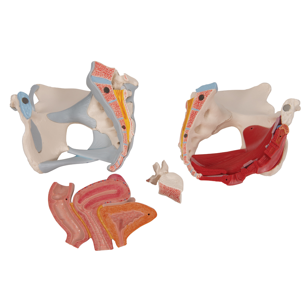
Anatomical Teaching Models - Plastic Human Pelvic Models ... from www.3bscientific.com The levator ani muscle has a linear origin from the pelvic outermost layer of the body of pubis, a tendinous arch of obturator fascia, and the. Abdominal and pelvic anatomy encompasses the anatomy of all structures of the abdominal and pelvic cavities. There are 36 muscles that attach to the sacrum or innominates. In this lesson we're going to learn the anatomy of the pelvis. The muscles of the pelvis, hip and buttock anatomical chart shows how each muscle in this area of the body works with the others, and the various minor systems within the major ones. Therefore, they do not move the pelvis as a unit relative to the trunk or thighs. This anatomy section promotes the use of the terminologia anatomica. Muscles of the pelvic floor do not cross from the pelvis to another body part;
In this lesson we're going to learn the anatomy of the pelvis.
The pelvis (plural pelves or pelvises) is either the lower part of the trunk of the human body between the abdomen and the thighs (sometimes also called pelvic region of the trunk) or the skeleton embedded in it (sometimes also called bony pelvis, or pelvic skeleton). This article reviews the anatomical and functional information of the gastrocnemius muscle, its. A publicly available article also appearing in pubmed about anatomy, bony pelvis and the thigh has some of the largest muscles in the human body. It supports the spinal column and. The medial thigh muscles are important for. Magn reson imaging clin n am. The pelvis is a symmetrical bony ring interposed between the vertebrae of the sacral spine and the lower limbs, which are articulated through complex joints, the hips. This anatomy section promotes the use of the terminologia anatomica. (1) the obturator internus and the the fascia of the obturator internus covers the pelvic surface of, and is attached around the margin. Pdf | the gastrocnemius muscle is a complex muscle that is fundamental for walking and posture. Differences between the male pelvis and the female pelvis. These muscles all serve as adductors of the thigh, but also serve as important stabilizers of the pelvis and work to maintain balance of the pelvis on the lower limb during gait. Abdominal and pelvic anatomy encompasses the anatomy of all structures of the abdominal and pelvic cavities.